detailed construcred images of neurons
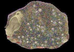
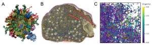
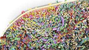
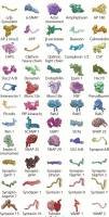


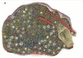
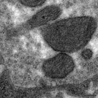
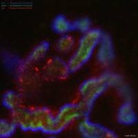
Models and visualizaiotns by Silvio Rizzoli
via book "How Life Works", Philip Ball
~
A snapshot of the contents of a cell. This image is from a computer model based on real microscope data from experimental studies of the varieties and numbers of copies of the various proteins in the cells in question, which are human neurons. Note that there will be other, smaller molecules and ions in the spaces between the proteins shown here. This part of the neuron sits at the synapse, the junction between one neuron and another. The molecules protruding from the cell surface help to transmit electrical signals across the gap. Image courtesy of Silvio Rizzoli, University Medical Centre Göttingen (Helm et al. 2021).
~
detailed pdf https://www.umg.eu/fileadmin/Redaktion/Dachportal/004_Forschung/id78_Science_Support/id81_Karriere_Foerderung/id337_Very_important_publications/GOE-VIP-4_Rizzoli.pdf
David Goodsell: Nanotransport ~ Synapse



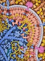
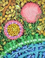
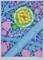
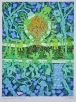
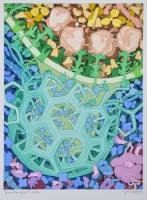
cool drawings of cell contents and inner workings
https://www.interaliamag.org/articles/david-goodsell/
via book "How Life Works", Philip Ball